If there is one thing that university faculty and administrators could do today to demonstrate their commitment to inclusion, not to mention teaching and learning over sorting and status, it would be to ban curve-based, norm-referenced grading. Many obstacles exist to the effective inclusion and success of students from underrepresented (and underserved) groups in science and related programs. Students and faculty often, and often correctly, perceive large introductory classes as “weed out” courses preferentially impacting underrepresented students. In the life sciences, many of these courses are “out-of-major” requirements, in which students find themselves taught with relatively little regard to the course’s relevance to bio-medical careers and interests. Often such out-of-major requirements spring not from a thoughtful decision by faculty as to their necessity, but because they are prerequisites for post-graduation admission to medical or graduate school. “In-major” instructors may not even explicitly incorporate or depend upon the materials taught in these out-0f-major courses – rare is the undergraduate molecular biology degree program that actually calls on students to use calculus or a working knowledge of physics, despite the fact that such skills may be relevant in certain biological contexts – see Magnetofiction – A Reader’s Guide. At the same time, those teaching “out of major” courses may overlook the fact that many (and sometimes most) of their students are non-chemistry, non-physics, and/or non-math majors. The result is that those teaching such classes fail to offer a doorway into the subject matter to any but those already comfortable with it. But reconsidering the design and relevance of these courses is no simple matter. Banning grading on a curve, on the other hand, can be implemented overnight (and by fiat if necessary).
So why ban grading on a curve? First and foremost, it would put faculty and institutions on record as valuing student learning outcomes (perhaps the best measure of effective teaching) over the sorting of students into easy-to-judge groups. Second, there simply is no pedagogical justification for curved grading, with the possible exception of providing a kludgy fix to correct for poorly designed examinations and courses. There are more than enough opportunities to sort students based on their motivation, talent, ambition, “grit,” and through the opportunities they seek after and successfully embraced (e.g., through volunteerism, internships, and independent study projects).
The negative impact of curving can be seen in a recent paper by Harris et al, (Reducing achievement gaps in undergraduate general chemistry …), who report a significant difference in overall student inclusion and subsequent success based on a small grade difference between a C, which allows a student to proceed with their studies (generally as successfully as those with higher grades) and a C-minus, which requires them to retake the course before proceeding (often driving them out of the major). Because Harris et al., analyzed curved courses, a subset of students cannot escape these effects. And poor grades disproportionately impact underrepresented and underserved groups – they say explicitly “you do not belong” rather than “how can I help you learn”.
Often naysayers disparage efforts to improve course design as “dumbing down” the course, rather than improving it. In many ways this is a situation analogous to blaming patients for getting sick or not responding to treatment, rather than conducting an objective analysis of the efficacy of the treatment. If medical practitioners had maintained this attitude, we would still be bleeding patients and accepting that more than a third are fated to die, rather than seeking effective treatments tailored to patients’ actual diseases – the basis of evidence-based medicine. We would have failed to develop antibiotics and vaccines – indeed, we would never have sought them out. Curving grades implies that course design and delivery are already optimal, and the fate of students is predetermined because only a percentage can possibly learn the material. It is, in an important sense, complacent quackery.
Banning grading on a curve, and labelling it for what it is – educational malpractice – would also change the dynamics of the classroom and might even foster an appreciation that a good teacher is one with the highest percentage of successful students, e.g. those who are retained in a degree program and graduate in a timely manner (hopefully within four years). Of course, such an alternative evaluation of teaching would reflect a department’s commitment to construct and deliver the most engaging, relevant, and effective educational program. Institutional resources might even be used to help departments generate more objective, instructor-independent evaluations of learning outcomes, in part to replace the current practice of student-based opinion surveys, which are often little more than measures of popularity. We might even see a revolution in which departments compete with one another to maximize student inclusion, retention, and outcomes (perhaps even to the extent of applying pressure on the design and delivery of “out of major” required courses offered by other departments).
“All a pipe dream” you might say, but the available data demonstrates that resources spent on rethinking course design, including engagement and relevance, can have significant effects on grades, retention, time to degree, and graduation rates. At the risk of being labeled as self-promoting, I offer the following to illustrate the possibilities: working with Melanie Cooper at Michigan State University, we have built such courses in general and organic chemistry and documented their impact, see Evaluating the extent of a large-scale transformation in gateway science courses.
Perhaps we should be encouraging students to seek out legal representation to hold institutions (and instructors) accountable for detrimental practices, such as grading on a curve. There might even come a time when professors and departments would find it prudent to purchase malpractice insurance if they insist on retaining and charging students for ineffective educational strategies.(1)
Acknowledgements: Thanks to daughter Rebecca who provided edits and legal references and Melanie Cooper who inspired the idea. Educate! image from theDorian De Long Arts & Music Scholarship site.
(1) One cannot help but wonder if such conduct could ever rise to the level of fraud. See, e.g., Bristol Bay Productions, LLC vs. Lampack, 312 P.3d 1155, 1160 (Colo. 2013) (“We have typically stated that a plaintiff seeking to prevail on a fraud claim must establish five elements: (1) that the defendant made a false representation of a material fact; (2) that the one making the representation knew it was false; (3) that the person to whom the representation was made was ignorant of the falsity; (4) that the representation was made with the intention that it be acted upon; and (5) that the reliance resulted in damage to the plaintiff.”).
Insights into student thinking & course design, part of the biofundamentals project.
Something that often eludes both instructors and instructional researchers is a clear appreciation of what it is that students do and do not know, what ideas they can and cannot call upon to solve problems and generate clear, coherent, and plausible explanations. What information – thought to have been presented effectively through past instruction, appears to be unavailable to students. As an example, few instructors would believe that students completing college level chemistry could possibly be confused about the differences between covalent and non-covalent molecular interactions, yet there is good evidence that they are (Williams et al., 2015). Unless these ideas, together with their conceptual bases and practical applications, are explicitly called out in the design and implementation of instructional materials, they often fail to become a working (relevant) part of the students’ conceptual tool-kit.
To identify ideas involved in understanding biological systems, we are using an upper division undergraduate course in developmental biology (blog link) to provide context; this is a final “capstone” junior/senior level course that comes after students have completed multiple required courses in chemistry and biology. Embryonic development integrates a range of molecular level processes, including the control of gene expression, cellular morphology and dynamics, through intrinsic and extrinsic signaling systems.
A key aspect of the course’s design is the use of formative assessment activities delivered through the beSocratic system. These activities generally include parts in which students are asked to draw a graph or diagram. Students are required to complete tasks before the start of each class meeting; their responses are used to inform in-class discussions, a situation akin to reviewing game film and coaching in sports. Analysis of student drawings and comments, carried out in collaboration with Melanie Cooper and her group at Michigan State University, can reveal unexpected aspects of students’ thinking (e.g. Williams et al., 2015). What emerges from this Socratic give and take is an improved appreciation of the qualities of the tasks that engage students (as well as those that do not), and insights into how students analyze specific tasks, what sets of ideas they see as necessary and which necessary ideas they ignore when generating explanatory and predictive models. Most importantly, they can reveal flaws in how necessary ideas are developed. While at an admittedly early stage in the project, here I sketch out some preliminary findings: the first of these deal with steady state concentration and response dynamics.
The ideas of steady state concentration and pathway dynamics were identified by Loertscher et al (2014)as two of five “threshold concepts” in biochemistry and presumably molecular biology as well. Given the non-equilibrium nature of biological systems, we consider the concentration of a particular molecule in a cell in dynamic terms, a function of its rate of synthesis (or importation from the environment) together with its rate of breakdown. On top of this dynamic, the activity of existing molecules can be regulated through various post-translational mechanisms. All of the populations of molecules within a cell or organism have a characteristic steady state concentration with the exception of genomic DNA, which while synthesized is not, in living organisms, degraded, although it is repaired.
In biological systems, molecules are often characterized by their “half life” but this can be confusing, since it is quite different from the way the term is used in physics, where students are likely to first be introduced to it.[1] Echos from physics can imply that a molecule’s half-life is an intrinsic feature of the molecule, rather than of the system in which the molecule finds itself. The equivalent of half-life would be doubling time, but these terms make sense only under specific conditions. In a system in which synthesis has stopped (synthesis rate = 0) the half life is the time it takes for the number of molecules in the system to decrease by 50%, while in the absence of degradation (degradation rate = 0), the doubling time is the time it takes to double the number of molecules in the system. Both degradation and synthesis rates are regulateable and can vary, often dramatically, in response to various stimuli.
In the case of RNA and polypeptide levels, the synthesis rate is determined by many distinct processes, including effective transcription factor concentrations, the signals that activate transcription factors, rates of binding of transcription factors to transcription factor binding sites (which can involve both DNA sequences and other proteins), as well as relevant binding affinities, and the rates associated with the recruitment and activation of DNA-dependent, RNA polymerase. Once activated, the rate of gene specific RNA synthesis will be influenced by the rate of RNA polymerization (nucleotide bases added per second) and the length of the RNA molecules synthesized. In eukaryotes, the newly formed RNA will generally need to have introns removed through interactions with splicing machinery, as well as other post-transcriptional reactions, after which the processed RNA will be transported from the nucleus to the cytoplasm through the nuclear pore complex. In the cytoplasm there are rates associated with the productive interaction of RNAs with the translational machinery (ribosomes and associated factors), and the rate at which polypeptide synthesis occurs (amino acids added per second) together with the length of the polypeptide synthesized (given that things are complicated enough, I will ignore processes such as those associated with the targeting of membrane proteins and codon usage, although these will be included in a new chapter in biofundamentals reasonably soon, I hope). On the degradative side, there are rates associated with interactions with nucleases (that breakdown RNAs) and proteinases (that breakdown polypeptides). These processes are energy requiring; generally driven by reactions coupled to the hydrolysis of adenosine triphosphate (ATP).
That these processes matter is illustrated nicely in work from Harima and colleagues (2014). The system, involved in the segmentation of the anterior region of the presomitic mesoderm, responds to signaling by activating the Hes7 gene, while the Hes7 gene product act to inhibit Hes7 gene expression. The result is an oscillatory response that is “tuned” by the length of the transcribed region (RNA length). This can be demonstrated experimentally by generating mice in which two of the genes three introns (Hes7-3) or all three introns (intron-less) are removed. Removing introns changes the oscillatory behavior of the system (Hes7 mRNA -blue and Hes7 protein – green)(Harima et al., 2013).
In the context of developmental biology, we use beSocratic activities to ask students to consider a molecule’s steady state concentration as a function of its synthesis and degradation rates, and to predict how the system would change when one or the other is altered. These ideas were presented in the context of observations by Schwanhausser et al (2011) that large discrepancies between steady state RNA and polypeptide concentrations are common and that there is an absence of a correlation between RNA and polypeptide half-lives (we also use these activities to introduce the general idea of correlation). In their responses, it was common to see students’ linking high steady state concentrations exclusively to long half-lives. Ask to consider the implications in terms of system responsiveness (in the specific context of a positively-acting transcription factor and target gene expression), students often presumed that a longer half-life would lead to higher steady state concentration which in turn would lead to increased target gene expression, primarily because collisions between the transcription factor and its DNA-binding sites would increase, leading to higher levels of target gene expression. This is an example of a p-prim (Hammer, 1996) – the heuristic that “more is more”, a presumption that is applicable to many systems.
In biological systems, however, this is generally not the case – responses “saturate”, that is increasing transcription factor concentration (or activity) above a certain level generally does not lead to a proportionate, or any increase in target gene expression. We would not call this a misconception, because this is an example of an idea that is useful in many situations, but generally isn’t in biological systems – where responses are generally inherently limited. The ubiquity and underlying mechanisms of response saturation need to be presented explicitly, and its impact on various processes reinforced repeatedly, preferably by having students use them to solve problems or construct plausible explanations. A related phenomenon that students seemed not to recognize involves the non-linearity of the initial response to a stimulus, in this case, the concentration of transcription factor below which target gene expression is not observed (or it may occur, but only transiently or within a few cells in the population, so as to be undetectable by the techniques used).
So what ideas do students need to call upon when they consider steady state concentration, how it changes, and the impact of such changes on system behavior? It seems we need to go beyond synthesis and degradation rates and include the molecular processes associated with setting the system’s response onset and saturation concentrations. First we need to help students appreciate why such behaviors (onset and saturation) occur – why doesn’t target gene expression begin as soon as a transcription factor appears in a cell? Why does gene expression level off when transcription factor concentrations rise above a certain level? The same questions apply to the types of threshold behaviors often associated with signaling systems. For example, in quorum sensing among unicellular organisms, the response of cells to the signal occurs over a limited concentration range, from off to full on. A related issue is associated with morphogen gradients (concentration gradients over space rather than time), in which there are multiple distinct types of “threshold” responses. One approach might be to develop a model in which we set the onset concentration close to the saturation concentration. The difficulty (or rather instructional challenge) here is that these are often complex processes involving cooperative as well as feedback interactions.
Our initial approach to steady state and thresholds has been to build activities based on the analysis of a regulatory network presented by Saka and Smith (2007), an analysis based on studies of early embryonic development in the frog Xenopus laevis. We chose the system because of its simplicity, involving only four components (although there are many other proteins associated with the actual system). Saka and Smith modeled the regulatory network controlling the expression of the transcription factor proteins Goosecoid (Gsc) and Brachyury (Xbra) in response to the secreted signaling protein activin (↓), a member of
the TGFβ superfamily of secreted signaling proteins (see Li and Elowitz, 2019). The network involves the positive action of Xbra on the gene encoding the transcription factor protein Xom. The system’s behavior depends on the values of various parameters, parameters that include response to activator (Activin), rates of synthesis and the half-lives of Gsc, Xbra, and Xom, and the degrees of regulatory cooperativity and responsiveness.
Depending upon these parameters, the system can produce a range of complex responses. In different regimes (→), increasing concentrations of activin (M) can lead, initially, to increasing, but mutually exclusive, expression of either Xba (B) or Gsc (A) as well as sharp transitions in which expression flips from one to the other, as Activin concentration increases, after which the response saturates. There are also conditions at very low Activin concentration (marked by ↑) in which both Xbra and Gsc are expressed at low levels, a situation that students are asked to explain.
Lessons learned: Based on their responses, captured through beSocratic and revealed during in class discussions, it appears that there is a need to be more explicit (early in the course, and perhaps the curriculum as well) when considering the mechanisms associated with response onset and saturation, in the context of how changes in the concentrations of regulatory factors (through changes in synthesis, turn-over, and activity) impact system responses. This may require a more quantitative approach to molecular dynamics and system behaviors. Here we may run into a problem, the often phobic responses of biology majors (and many faculty) to mathematical analyses. Even the simplest of models, such as that of Saka and Smith, require a consideration of factors generally unfamiliar to students, concepts and skills that may well not be emphasized or mastered in prerequisite courses. The trick is to define realistic, attainable, and non-trivial goals – we are certainly not going to succeed in getting late stage molecular biology students with rudimentary math skills to solve systems of differential equations in a developmental biology course. But perhaps we can build up the instincts needed to appreciate the molecular processes involved in the behavior of systems whose behavior evolves overtime in response to various external signals (which is, of course, pretty much every biological system).
Footnotes:
[1] A similar situation exists in the context of the term “spontaneous” in chemistry and biology. In chemistry spontaneous means thermodynamically favorable, while in standard usage (and generally in biology) spontaneous implies that a reaction is proceeding at a measurable, functionally significant rate. Yet another insight that emerged through discussions with Melanie Cooper.
Mike Klymkowsky
Literature cited
Hammer, D. (1996). Misconceptions or p-prims. How might alternative perspectives of cognitive structure influence instructional perceptions and intentions. Journal of the Learning Sciences5, 97-127.
Harima, Y., Imayoshi, I., Shimojo, H., Kobayashi, T. and Kageyama, R. (2014). The roles and mechanism of ultradian oscillatory expression of the mouse Hes genes. In Seminars in cell & developmental biology, pp. 85-90: Elsevier.
Harima, Y., Takashima, Y., Ueda, Y., Ohtsuka, T. and Kageyama, R. (2013). Accelerating the tempo of the segmentation clock by reducing the number of introns in the Hes7 gene. Cell Reports3, 1-7.
Li, P. and Elowitz, M. B. (2019). Communication codes in developmental signaling pathways. Development146, dev170977.
Loertscher, J., Green, D., Lewis, J. E., Lin, S. and Minderhout, V. (2014). Identification of threshold concepts for biochemistry. CBE—Life Sciences Education 13, 516-528.
Saka, Y. and Smith, J. C. (2007). A mechanism for the sharp transition of morphogen gradient interpretation in Xenopus. BMC Dev Biol7, 47.
Schwanhäusser, B., Busse, D., Li, N., Dittmar, G., Schuchhardt, J., Wolf, J., Chen, W. and Selbach, M. (2011). Global quantification of mammalian gene expression control. Nature 473, 337.
Williams, L. C., Underwood, S. M., Klymkowsky, M. W. and Cooper, M. M. (2015). Are Noncovalent Interactions an Achilles Heel in Chemistry Education? A Comparison of Instructional Approaches. Journal of Chemical Education92, 1979–1987.
Please note, given the move from PLoS some of the links in the posts may be broken; some minor editing in process. All by Mike Klymkowsky unless otherwise noted
Embryogenesis is based on a framework of social (cell-cell) interactions, initial and early asymmetries, and cascading cell-cell signaling and gene regulatory networks (DEVO posts one, two, & three). The result is the generation of embryonic axes, germ layers (ectoderm, mesoderm, endoderm), various organs and tissues (brains, limbs, kidneys, hearts, and such) and their characteristic cell types, their patterning, and their coordination into a functioning organism. It is well established that all animals share a common ancestor (hundreds of millions of years ago) and that a number of molecularmodules were already present in that common ancestor.
At the same time evolutionary processes are, and need to be, flexible enough to generate the great diversity of organisms, with their various adaptations to particular life-styles. The extent of both conservation and flexibility (new genes, new mechanisms) in developmental systems is, however, surprising. Perhaps the most striking evidence for the depth of this conservation was supplied by the discovery of the organization of the Hox gene cluster in the fruit fly Drosophila and in the mouse (and other vertebrates). In both, the Hox genes are arranged and expressed in a common genomic and expression patterns. But as noted by Denis Duboule (2007) Hox gene organization is often presented in textbooks in a distorted manner (↓).
The Hox gene clusters of vertebrates are compact, but are split, disorganized, and even “atomized” in other types of organisms. Similarly, processes that might appear foundational, such as the role of the Bicoid gradient in the early fruit fly embryo (a standard topic in developmental biology textbooks), is in fact restricted to a small subset of flies (Stauber et al., 1999). New genes can be generated through well defined processes, such as gene duplication and divergence, or they can arise de novo out of sequence noise (Carvunis et al., 2012; Zhao et al., 2014 – see Van Oss & Carvunis 2019. De novo gene birth). Comparative genomic analyses can reveal the origins of specific adaptations (see Stauber et al., 1999).The result is that organisms as closely related to each other as the great apes (including humans) have significant species-specific genetic differences (see Florio et al., 2018; McLean et al., 2011; Sassa, 2013 and references therein) as well as common molecular and cellular mechanisms.
A universal (?) feature of developing systems – gradients and non-linear responses: There is a predilection to find (and even more to teach) simple mechanisms that attempt to explain everything (witness the distortion of the Hox cluster, above) – a form of physics “theory of everything” envy.But the historic nature, evolutionary plasticity, and need for regulatory robustness generally lead to complex and idiosyncratic responses in biological systems.Biological systems are not “intelligently designed” but rather cobbled together over time through noise (mutation) and selection (Jacob, 1977)(see blog post).
That said, acommon (universal?) developmental process appears to be the transformation of asymmetries into unambiguous cell fate decisions. Such responses are based on threshold events controlled by a range of molecular behaviors, leading to discrete gene expression states. We can approach the question of how such decisions are made from both an abstract and a concrete perspective. Here I outline my initial approach – I plan to introduce organism specific details as needed.I start with the response to a signaling gradient, such as that found in many developmental systems, including the vertebrate spinal cord (top image Briscoe and Small, 2015) and the early Drosophila embryo (Lipshitz, 2009)(↓).
We begin with a gradient in the concentration of a “regulatory molecule” (the regulator).The shape of the gradient depends upon the sites and rates of synthesis, transport away from these sites, and turnover (degradation and/or inactivation). We assume, for simplicity’s sake, that the regulator directly controls the expression of target gene(s). Such a molecule binds in a sequence specific manner to regulatory sites, there could be a few or hundreds, and lead to the activation (or inhibition) of the DNA-dependent, RNA polymerase (polymerase), which generates RNA molecules complementary to one strand of the DNA. Both the binding of the regulator and the polymerase are stochastic processes, driven by diffusion, molecular collisions, and binding interactions.(1)
Let us now consider the response of target gene(s) as a function of cell position within the gradient. We might (naively) expect that the rate of target gene expression would be a simple function of regulator concentration. For an activator, where the gradient is high, target gene expression would be high, where the gradient concentration is low, target gene expression would be low – in between, target gene expression would be proportional to regulator concentration.But generally we find something different, we find that the expression of target genes is non-uniform, that is there are thresholds in the gradient: on one side of the threshold concentration the target gene is completely off (not expressed), while on the other side of the threshold concentration, the target gene is fully on (maximally expressed).The target gene responds as if it is controlled by an on-off switch. How do we understand the molecular basis for this behavior?
Distinct mechanisms are used in different systems, but we will consider a system from the gastrointestinal bacteria E. coli that students may already be familiar with; these are the genes that enable E. coli to digest the mammalian milk sugar lactose.They encode a protein needed to importlactose into a bacterial cell and an enzyme needed to break lactose down so that it can be metabolized.Given the energetic cost to synthesize these proteins, it is in the bacterium’s adaptive self interest to synthesize them only when lactose is present at sufficient concentrations in their environment.The response is functionally similar to that associated with quorum sensing, which is also governed by threshold effects. Similarly cells respond to the concentration of regulator molecules (in a gradient) by turning on specific genes in specific domains, rather than uniformly.
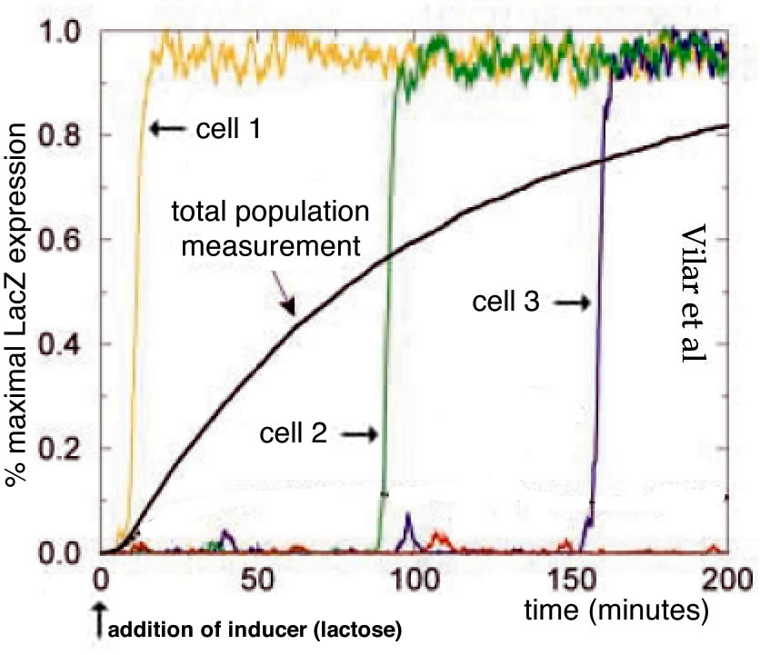
Now let us look in a little more detail at the behavior of the lactose utilization system in E. coli following an analysis by Vilar et al (2003)(2).At an extracellular lactose concentration below the threshold, the system is off.If we increase the extracellular lactose concentration above threshold the system turns on, the lactose permease and β-galactosidase proteins are made and lactose can enter the cell and be broken down to produce metabolizable sugars.By looking at individual cells, we find that they transition, apparently stochastically from off to on (→), but whether they stay on depends upon the extracellular lactose concentration. We can define a concentration, the maintenance concentration, below the threshold, at which “on” cells will remain on, while “off” cells will remain off.
The circuitry of the lactose system is well defined(Jacob and Monod, 1961; Lewis, 2013; Monod et al., 1963)(↓).The lacI gene encodes the lactose operon repressor protein and it is expressed constituately at a low level; it binds to sequences in the lac operon and inhibits transcription.The lac operon itself contains three genes whose expression is regulated by a constituatively active promoter.LacY encodes the permease while the lacZ encodes β-galactosidase.β-galactosidase has two functions: it catalyzes the reaction that transforms lactose into allolactone and it cleaves lactose into the metabolically useful sugars glucose and galactose. Allolactone is an allosteric modulator of the Lac repressor protein; if allolactone is present, it binds to lac epressor proteins and inactivates them, allowing lac operon expression.
The cell normally contains only ~10 lactose repressor proteins. Periodically (stochastically), even in the absence of lactose, and so its derivative allolactone, the lac operon promoter region is free of repressor proteins, and a lactose operon is briefly expressed – a few LacY and LacZpolypeptides are synthesized (↓).This noisy leakiness in the regulation of the lac operon allows the cell to respond if lactose happens to be present – some lactose molecules enter the cell through the permease, are converted to allolactone by β-galactosidase.Allolactone is an allosteric effector of the lac repressor; when present it binds to and inactivates the lac repressor protein so that it no longer binds to its target sequences (the operator or “O” sites).In the absence of repressor binding, the lac operon is expressed.If lactose is not present, the lac operon is inhibited and lacY and LacZ disappear from the cell by turnover or growth associated dilution.
The question of how the threshold concentration for various signal-regulated decisions is set often involves homeostatic processes that oppose the signaling response. The binding and activation of regulators can involve cooperative interactions between molecular components and both positive and negative feedback effects.
In the case of patterning a tissue, in terms of regional responses to a signaling gradient, there can be multiple regulatory thresholds for different genes, as well as indirect effects, where the initiation of gene expression of one set of target genes impacts the sensitive expression of subsequent sets of genes.One widely noted mechanism, known as reaction-diffusion, was suggested by the English mathematician Alan Turing (see Kondo and Miura, 2010) – it postulates a two component system. One component is an activator of gene expression, which in addition to its own various targets, positively regulates its own expression. The second component is a repressor of the first.Both of these two regulator molecules are released by the signaling cell or cells; the repressor diffuses away from the source faster than the activator does.The result can be a domain of target gene expression (where the concentration of activator is sufficient to escape repression), surrounded by a zone in which expression is inhibited (where repressor concentration is sufficient to inhibit the activator).Depending upon the geometry of the system, this can result in discrete regions (dots or stripes) of primary target gene expression (see Sheth et al., 2012).In real systems there are often multiple gradients present; their relative orientations can produce a range of patterns.
The point of all of this, is that when we approach a particular system – we need to consider the mechanisms involved.Typically they are selected to produce desired phenotypes, but also to be robust in the sense that they need to produce the same patterns even if the system in which they occur is subject to perturbations, such as embryo/tissue size (due to differences in cell division / growth rates) and temperature and other environmental variables.
note: figures returned – updated 13 November 2020.
Footnotes:
While stochastic (random) these processes can still be predictable.A classic example involves the decay of an unstable isotope (atom), which is predictable at the population level, but unpredictable at the level of an individual atom.Similarly, in biological systems, the binding and unbinding of molecules to one another, such as a protein transcription regulator to its target DNA sequence is stochastic but can be predictable in a large enough population.
Briscoe & Small (2015). Morphogen rules: design principles of gradient-mediated embryo patterning. Development 142, 3996-4009.
Carvunis et al (2012). Proto-genes and de novo gene birth. Nature 487, 370.
Duboule (2007). The rise and fall of Hox gene clusters. Development 134, 2549-2560.
Florio et al (2018). Evolution and cell-type specificity of human-specific genes preferentially expressed in progenitors of fetal neocortex. eLife 7.
Jacob (1977). Evolution and tinkering. Science 196, 1161-1166.
Jacob & Monod (1961). Genetic regulatory mechanisms in the synthesis of proteins. Journal of Molecular Biology 3, 318-356.
Kondo & Miura (2010). Reaction-diffusion model as a framework for understanding biological pattern formation. Science 329, 1616-1620.
Lewis (2013). Allostery and the lac Operon. Journal of Molecular Biology 425, 2309-2316.
Lipshitz (2009). Follow the mRNA: a new model for Bicoid gradient formation. Nature Reviews Molecular Cell Biology 10, 509.
McLean et al (2011). Human-specific loss of regulatory DNA and the evolution of human-specific traits. Nature 471, 216-219.
Monod Changeux & Jacob (1963). Allosteric proteins and cellular control systems. Journal of Molecular Biology 6, 306-329.
Sassa (2013). The role of human-specific gene duplications during brain development and evolution. Journal of Neurogenetics 27, 86-96.
Sheth et al (2012). Hox genes regulate digit patterning by controlling the wavelength of a Turing-type mechanism. Science 338, 1476-1480.
Stauber et al (1999). The anterior determinant bicoid of Drosophila is a derived Hox class 3 gene. Proceedings of the National Academy of Sciences 96, 3786-3789.
Vilar et al (2003). Modeling network dynamics: the lac operon, a case study. J Cell Biol 161, 471-476.
21st Century DEVO-2 In the first post in this series [link], I introduced the observation that single celled organisms can change their behaviors, often in response to social signals.They can respond to changing environments and can differentiate from one cellular state to the another. Differentiation involves changes in which sets of genes are expressed, which polypeptides and proteins are made [previous post], where the proteins end up within the cell, and which behaviors are displayed by the organism. Differentiation enables individuals to adapt to hostile conditions and to exploit various opportunities.
The ability of individuals to cooperate with one another, through processes such as quorum sensing, enables them to tune their responses so that they are appropriate and useful. Social interactions also makes it possible for them to produce behaviors that would be difficult or impossible for isolated individuals.Once individual organisms learn, evolutionarily, how to cooperate, new opportunities and challenges (cheaters) emerge. There are strategies that can enable an organism to adapt to a wider range of environments, or to become highly specialized to a specific environment,through the production of increasingly complex behaviors.As described previously, many of these cooperative strategies can be adopted by single celled organisms, but others require a level of multicellularity.Multicellularity can be transient – a pragmatic response to specific conditions, or it can be (if we ignore the short time that gametes exist as single cells) permanent, allowing the organism to develop the range of specialized cells types needed to build large, macroscopic organisms with complex and coordinated behaviors. In appears that various forms of multicellularity have arisen independently in a range of lineages (Bonner, 1998; Knoll, 2011). We can divide multicellularity into two distinct types, aggregative and clonal – which we will discuss in turn (1). Aggregative (transient) multicellularity:Once organisms had developed quorum sensing, they can monitor the density of related organisms in their environment and turn or (or off) specific genes (or sets of genes, necessary to produce a specific behavior.While there are many variants, one model for sucha behavior isa genetic toggle switch, in which a particular gene (or genes) can be switched on or off in response to environmental signals acting as allosteric regulators of transcription factor proteins (see Gardner et al., 2000).Here is an example of an activity (↓) that we will consider in class to assess our understanding of the molecular processes involved.
One outcome of such a signaling system is to provoke the directional migration of amoeba and their aggregation to form the transient multicellular “slug”.Such behaviors has been observedin a range of normally unicellular organisms (see Hillmann et al., 2018)(↓). The classic example isthe cellular slime mold Dictyostelium discoideum (Loomis, 2014).Under normal conditions, these
unicellular amoeboid eukaryotes migrate, eating bacteria and such. In this state, the range of an individual’s movement is restricted to short distances.However when conditions turn hostile, specifically a lack of necessary nitrogen compounds, there is a compelling reason to abandon one environment and migrate to another, more distant that a single-celled organism could reach. This is a behavior that depends upon the presence of a sufficient density (cells/unit volume) of cells that enables them to: 1) recognize one another’s presence (through quorum sensing), 2) find each other through directed (chemotactic) migration, and 3) form a multicellular slug that can go on to differentiate. Upon differentiation about 20% of the cells differentiate (and die), forming a stalk that lifts the other ~80% of the cells into the air.These non-stalk cells (the survivors) differentiate into spore (resistant to drying out) cells that are released into the air where they can be carried to new locations, establishing new populations.
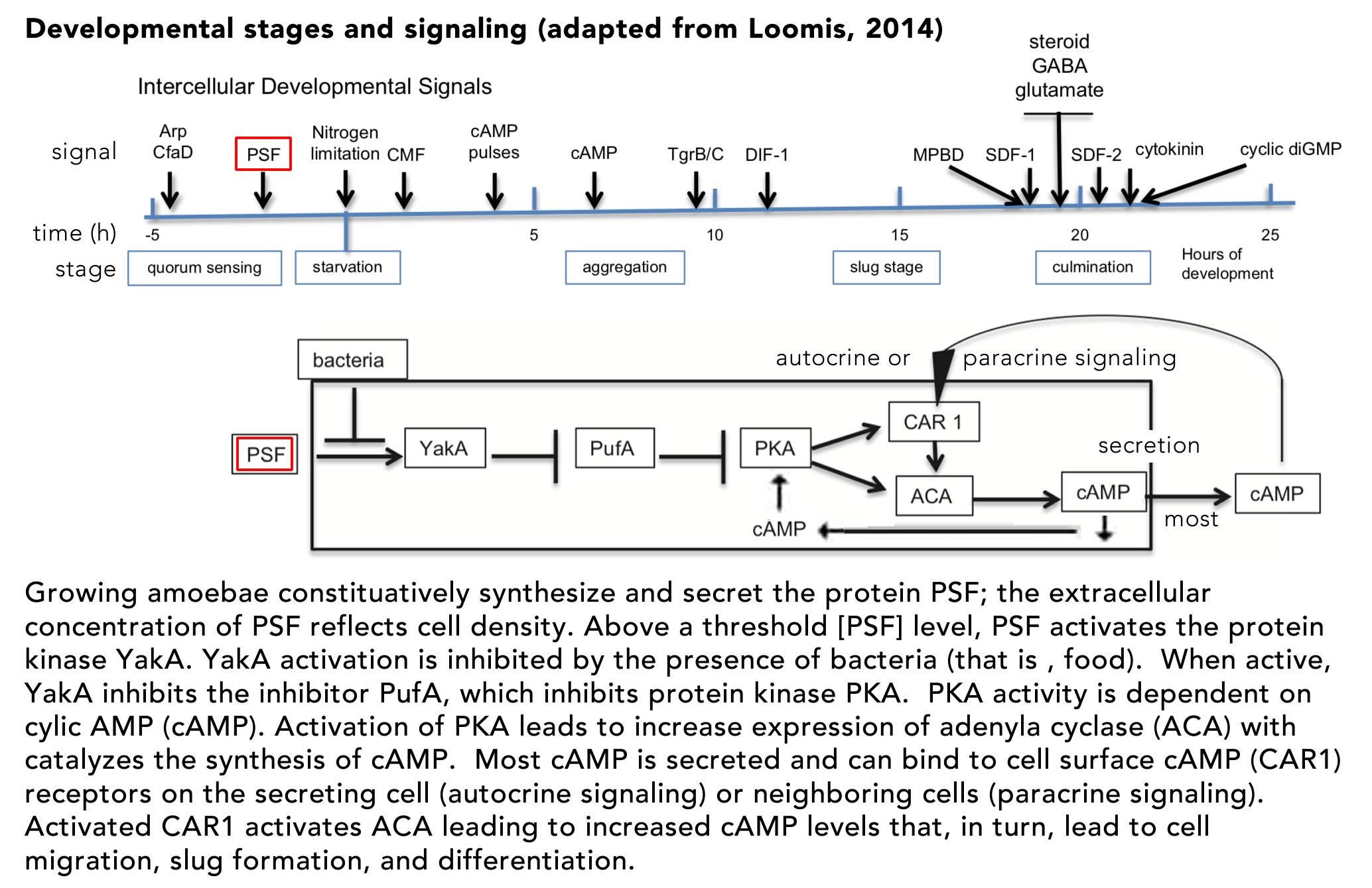
The process of cellular differentiation in D. discoideum has been worked out in molecular detail and involves two distinct signaling systems: the secreted pre-starvation factor (PSF) protein and cyclic AMP (cAMP).PSF is a quorum signaling protein that also serves to activate the cell aggregation and differentiation program (FIG. ↓)
If bacteria, that is food, are present, the activity of PSF is inhibited andcells remain in their single cell state. The key regulator of downstream aggregation and differentiation is the cAMP-dependent protein kinase PKA.In the unicellular state, PKA activity is inhibited by PufA.As PSF increases, while food levels decrease, YakA activity increases, inactivating PufA, leading to increased PKA activity.Active PKA induces the synthesis of two downstream proteins, adenylate cyclase (ACA) and the cAMP receptor (CAR1). ACA catalyzes cAMP synthesis, much of which is secreted from the cell as a signaling molecule. The membrane-bound CAR1 protein acts as a receptor for autocrine (on the cAMP secreting cell) and paracrine (on neighboring cells) signaling.The binding of cAMP to CAR1 leads to further activation of PKA, increasing cAMP synthesis and secretion – a positive feed-back loop. As cAMP levels increase, downstream genes are activated (and inhibited) leading cells to migrate toward one another, their adhesion to form a slug.Once the slug forms and migrates to an appropriate site, the process of differentiation (and death) leading to stalk and spore formation begins. The fates of the aggregated cells is determined stochastically, but social cheaters can arise. Mutations can lead to individuals that avoid becoming stalk cells.In the long run, if all individuals were to become cheaters, it would be impossible to form a stalk, so the purpose of social cooperation would be impossible to achieve.In the face of environmental variation, populations invaded by cheaters are more likely to become extinct.For our purposes the various defenses against cheaters are best left to other courses (see here if interested Strassmann et al., 2000).
Clonal (permanent) multicellularity:The type of multicellularity that most developmental biology courses focus on is what is termed clonal multicellularity – the organism is a clone of an original cell, the zygote, a diploid cell produced by the fusion of sperm and egg, haploid cells formed through the process of meiosis (2).It is during meiosis that most basic genetic processes occur, that is the recombination between maternal and paternal chromosomes leading to the shuffling of alleles along a chromosome, and the independent segregation of chromosomes to form haploid gametes, gametes that are genetically distinct from those present in either parent. Once the zygote forms, subsequent cell divisions involve mitosis, with only a subset of differentiated cells, the cells of the germ line, capable of entering meiosis.
Non-germ line, that is somatic cells, grow and divide. They interact with one another directly and through various signaling processes to produce cells with distinct patterns of gene expression, and so differentiated behaviors.A key difference from a unicellular organism, is that the cells will (largely) stay attached to one another, or to extracellular matrix materials secreted by themselves and their neighbors.The result is ensembles of cells displaying different specializations and behaviors.As such cellular colonies get larger, they face a number of physical constraints – for example, cells are open non-equilibrium systems, to maintain themselves and to grow and reproduce, they need to import matter and energy from the external world. Cells also produce a range of, often toxic, waste products that need to be removed.As the cluster of zygote-derived cells grows larger, and includes more and more cells, some cells will become internal and so cut off from necessary resources. While diffusive processes are often adequate when a cell is bathed in an aqueous solution, they are inadequate for a cell in the interior of a large cell aggregate (3).The limits of diffusive processes necessitate other strategies for resource delivery and waste removal; this includes the formation of tubular vascular systems (such as capillaries, arteries, veins) and contractile systems (hearts and such) to pump fluids through these vessels, as well as cells specialized to process and transport a range of nutrients (such as blood cells).As organisms get larger, their movements require contractile machines (muscle, cartilage, tendons, bones, etc) driving tails, fins, legs, wings, etc. The coordination of such motile systems involves neurons, ganglia, and brains. There is also a need to establish barriers between the insides of an organism and the outside world (skin, pulmonary, and gastrointestinal linings) and the need to protect the interior environment from invading pathogens (the immune system).The process of developing these various systems depends upon controlling patterns of cell growth, division, and specialization (consider the formation of an arm), as well as the controlled elimination of cells (apoptosis), important in morphogenesis (forming fingers from paddle-shaped appendages), the maturation of the immune system (eliminating cells that react against self), and the wiring up, and adaptation of the nervous system. Such changes are analogous to those involved in aggregative multicellularity.
Origins of multicellularity:While aggregative multicellularity involves an extension of quorum sensing and social cooperation between genetically distinct, but related individuals, we can wonder whether similar drivers are responsible for clonal multicellularity.There are a number of imaginable adaptive (evolutionary) drivers but two spring to mind: a way to avoid predators by getting bigger than the predators and as a way to produce varied structures needed to exploit various ecological niches and life styles. An example of the first type of driver of multicellularity is offered by the studies of Boraas et al(1998). They cultured the unicellular green alga Chlorella vulgaris, together with a unicellular predator, the phagotrophic flagellated protist Ochromonas vallescia. After less than 100 generations (cell divisions), they observed the appearance of multicellular, and presumable inedible (or at least less easily edible), forms. Once selected, this trait appears to be stable, such that “colonies retained the eight-celled form indefinitely in continuous culture”.To my knowledge, the genetic basis for this multicellularity remains to be determined.
Cell Differentiation:One feature of simple colonial organisms is that when dissociated into individual cells, each cell is capable of regenerating a new organism. The presence of multiple (closely related) cells in a single colony opens up the possibility of social interactions; this is distinct from the case in aggregative multicellularity, where social cooperation came first. Social cooperation within a clonal metazoan means that most cells “give up” their ability to reproduce a new organism (a process involving meiosis). Such irreversible social interactions mark the transition from a colonial organism to a true multicellular organism. As social integration increases, cells can differentiate so as to perform increasingly specialized functions, functions incompatible with cell division. Think for a moment about a human neuron or skeletal muscle cell – in both cases, cell division is no longer possible (apparently). Nevertheless, the normal functioning of such cells enhances the reproductive success of the organism as a whole – a classic example of inclusive fitness (remember heterocysts?)Modern techniques of single cell sequencing and data analysis have now been employed to map this process of cellular differentiation in increasingly great detail, observations that will inform our later discussions (see Briggs et al., 2018 and future posts). In contrast, the unregulated growth of a cancer cell is an example of an asocial behavior, an asocial behavior that is ultimately futile, except in those rare cases (four known at this point) in which a cancer cell can move from one organism to another (Ujvari et al., 2016).
Unicellular affordances for multicellularity:When considering the design of a developmental biology course, we are faced with the diversity of living organisms – the basic observation that Darwin, Wallace, their progenitors and disciplinary descendants set out to solve. After all there are many millions of different types of organisms; among the multicellular eukaryotes, there are six major group : the ascomycetes and basidiomycetes fungi, the florideophyte red algae, laminarialean brown algae, embryophytic land plants and animals
(Knoll, 2011 ↑).Our focus will be on animals. “All members of Animalia are multicellular, and all are heterotrophs (i.e., they rely directly or indirectly on other organisms for their nourishment). Most ingest food and digest it in an internal cavity.” [Mayer link].From a macroscopic perspective, most animals have (or had at one time during their development) an anterior to posterior, that is head to tail, axis. Those that can crawl, swim, walk, or fly typically have a dorsal-ventral or back to belly axis, and some have a left-right axis as well.
But to be clear, a discussion of the various types of animals is well beyond the scope of any introductory course in developmental biology, in part because there are 35 (assuming no more are discovered) different “types” (phyla) of animals – nicely illustrated at this website [BBC: 35 types of animals, most of whom are really weird)].So again, our primary focus will be on one group, the vertebrates – humans are members of this group.We will also consider experimental insights derived from studies of various “model” systems, including organisms from another metazoan group, theecdysozoa (organisms that shed their outer layer as they grow bigger), a group that includes fruit flies and nematode worms.
My goal will be to ignore most of the specialized terminology found in the scholarly literature, which can rapidly turn a biology course into a vocabulary lesson and that add little to understanding of basic processes relevant to a general understanding of developmental processes (and relevant to human biology, medicine, and biotechnology). This approach is made possible by the discovery that the basic processes associated with animal (and metazoan) development are conserved. In this light, no observation has been more impactful than the discovery that the nature and organization of the genes involved in specifying the head to tail axes of the fruit fly and vertebrates (such as the mouse and human) is extremely similar in terms of genomic organization and function (Lappin et al., 2006 ↓), an observation that we will return to repeatedly.Such molecular similarities extend to cell-cell and cell-matrix adhesion systems, systems that release and respond to various signaling molecules, controlling cell behavior and gene expression, and reflects the evolutionary conservation and the common ancestry of all animals (Brunet and King, 2017; Knoll, 2011).
What can we know about the common ancestor of the animals?Early on in the history of comparative cellular anatomy, the striking structural similarities between the feeding system of choanoflagellate protozoans, a motile (microtubule-based) flagellum a surrounded by a “collar”of microfilament-based microvilli) and a structurally similar organelle in a range of multicellular organisms led to the suggestion that choanoflagellates and animals shared a common ancestor.The advent of genomic sequencing and analysis has only strengthened this hypothesis, namely that choanoflagellates and animals form a unified evolutionary clade, the ‘Choanozoa’(see tree↑ above)(Brunet and King, 2017).Moreover, “many genes required for animal multicellularity (e.g., tyrosine kinases, cadherins, integrins, and extracellular matrix domains) evolved before animal origins”.The implications is that the Choanozoan ancestor was predisposed to exploit some of the early opportunities offered by clonal multicellularity. These pre-existing affordances, together with newly arising genes and proteins (Long et al., 2013) were exploited in multiple lineages in the generation of multicellular organisms (see Knoll, 2011).
Basically to understand what happened next, some ~600 million years ago or so, we will approach the various processes involved in the shaping of animal development.Because all types of developmental processes, including the unicellular to colonial transition, involve changes in gene expression, we will begin with the factors involved in the regulation of gene expression.
Footnotes: 1). Please excuse the inclusive plural, but it seems appropriate in the context of what I hope will be a highly interactive course.
2). I will explicitly ignore variants as (largely) distractions, better suited for more highly specialized courses. 3). We will return to this problem when (late in the course, I think) we will discuss the properties of induced pluripotent stem cell (iPSC) derived organoids.
Literature cited: Bonner, J. T. (1998). The origins of multicellularity. Integrative Biology: Issues, News, and Reviews: Published in Association with The Society for Integrative and Comparative Biology 1, 27-36.
Boraas, M. E., Seale, D. B. and Boxhorn, J. E. (1998). Phagotrophy by a flagellate selects for colonial prey: a possible origin of multicellularity. Evolutionary Ecology 12, 153-164.
Briggs, J. A., Weinreb, C., Wagner, D. E., Megason, S., Peshkin, L., Kirschner, M. W. and Klein, A. M. (2018). The dynamics of gene expression in vertebrate embryogenesis at single-cell resolution. Science 360, eaar5780.
Brunet, T. and King, N. (2017). The origin of animal multicellularity and cell differentiation. Developmental cell 43, 124-140.
Gardner, T. S., Cantor, C. R. and Collins, J. J. (2000). Construction of a genetic toggle switch in Escherichia coli. Nature 403, 339-342.
Hillmann, F., Forbes, G., Novohradská, S., Ferling, I., Riege, K., Groth, M., Westermann, M., Marz, M., Spaller, T. and Winckler, T. (2018). Multiple roots of fruiting body formation in Amoebozoa. Genome biology and evolution 10, 591-606.
Knoll, A. H. (2011). The multiple origins of complex multicellularity. Annual Review of Earth and Planetary Sciences 39, 217-239.
Lappin, T. R., Grier, D. G., Thompson, A. and Halliday, H. L. (2006). HOX genes: seductive science, mysterious mechanisms. The Ulster medical journal 75, 23.
Long, M., VanKuren, N. W., Chen, S. and Vibranovski, M. D. (2013). New gene evolution: little did we know. Annual review of genetics 47, 307-333.
Loomis, W. F. (2014). Cell signaling during development of Dictyostelium. Developmental biology 391, 1-16.
Strassmann, J. E., Zhu, Y. and Queller, D. C. (2000). Altruism and social cheating in the social amoeba Dictyostelium discoideum. Nature 408, 965-967.
Ujvari, B., Gatenby, R. A. and Thomas, F. (2016). Transmissible cancers, are they more common than thought? Evolutionary applications 9, 633-634.