Molecular machines and the place of physics in the biology curriculum

The other day, through no fault of my own, I found myself looking at the courses required by our molecular biology undergraduate degree program. I discovered a requirement for a 5 credit hour physics course, and a recommendation that this course be taken in the students’ senior year – a point in their studies when most have already completed their required biology courses.  Befuddlement struck me, what was the point of requiring an introductory physics course in the context of a molecular biology major?  Was this an example of time-travel (via wormholes or some other esoteric imagining) in which a physics course in the future impacts a students’ understanding of molecular biology in the past?  I was also struck by the possibility that requiring such a course in the students’ senior year would measurably impact their time to degree. 

In a search for clarity and possible enlightenment, I reflected back on my own experiences in an undergraduate biophysics degree program – as a practicing cell and molecular  biologist, I was somewhat confused. I could not put my finger on the purpose of our physics requirement, except perhaps the admirable goal of supporting physics graduate students. But then, after feverish reflections on the responsibilities of faculty in the design of the courses and curricula they prescribe for their students and the more general concepts of instructional (best) practice and malpractice, my mind calmed, perhaps because I was distracted by an article on Oxford Nanopore’s MinION (↓), a “portable real-time device for DNA and RNA sequencing”,a device that plugs into the USB port on one’s laptop!

Distracted from the potentially quixotic problem of how to achieve effective educational reform at the undergraduate level, I found myself driven on by an insatiable curiosity (or a deep-seated insecurity) to insure that I actually understood how this latest generation of DNA sequencers worked. This led me to a paper by Meni Wanunu (2012. Nanopores: A journey towards DNA sequencing)[1].  On reading the paper, I found myself returning to my original belief, yes, understanding physics is critical to developing a molecular-level understanding of how biological systems work, BUT it was just not the physics normally inflicted upon (required of) students [2]. Certainly this was no new idea.  Bruce Alberts had written on this topic a number of times, most dramatically in his 1989 paper “The cell as a collection of molecular machines” [3].  Rather sadly, and not withstanding much handwringing about the importance of expanding student interest in, and understanding of, STEM disciplines, not much of substance in this area has occurred. While (some minority of) physics courses may have adopted active engagement pedagogies (in the meaning of Hake [4]) most insist on teaching macroscopic physics, rather than to focus on, or even to consider, the molecular level physics relevant to biological systems, explicitly the physics of protein machines in a cellular (biological) context. Why sadly, because conventional, that is non-biologically relevant introductory physics and chemistry courses, all to often serve the role of a hazing ritual, driving many students out of biology-based careers [5], in part I suspect, because they often seem irrelevant to students’ interests in the workings of biological systems. (footnote 1)  

Nanopore’s sequencer and Wanunu’s article (footnote 2) got me thinking again about biological machines, of which there are a great number, ranging from pumps, propellers, and oars to  various types of transporters, molecular truckers that move chromosomes, membrane vesicles, and parts of cells with respect to one another, to DNA detanglers, protein unfolders, and molecular recyclers (↓). 

Nanopore’s sequencer works based on the fact that as a single strand of DNA (or RNA) moves through a narrow pore, the different bases (A,C,T,G) occlude the pore to different extents, allowing different numbers of ions, different amounts of current, to pass through the pore. These current differences can be detected, and allows for a nucleotide sequence to be “read” as the nucleic acid strand moves through the pore. Understanding the process involves understanding how molecules move, that is the physics of molecular collisions and energy transfer, how proteins and membranes allow and restrict ion movement, and the impact of chemical gradients and electrical fields across a membrane on molecular movements  – all physical concepts of widespread significance in biological systems (here is an example of where a better understanding of physics could be useful to biologists).  Such ideas can be extended to the more general questions of how molecules move within the cell, and the effects of molecular size and inter-molecular interactions within a concentrated solution of proteins, protein polymers, lipid membranes, and nucleic acids, such as described in Oliverira et al., (2016 Increased cytoplasmic viscosity hampers aggregate polar segregation in Escherichia coli)[6].  At the molecular level, the processes, while biased by electric fields (potentials) and concentration gradients, are stochastic (noisy). Understanding of stochastic processes is difficult for students [7], but critical to developing an appreciation of how such processes can lead to phenotypic  differences between cells with the same genotypes (previous post) and how such noisy processes are managed by the cell and within a multicellular organism.   

As path leads on to path, I found myself considering the (←) spear-chucking protein machine present in the pathogenic bacteria Vibrio cholerae; this molecular machine is used to inject toxins into neighbors that the bacterium happens to bump into (see Joshi et al., 2017. Rules of Engagement: The Type VI Secretion System in Vibrio cholerae)[8].  The system is complex and acts much like a spring-loaded and rather “inhumane” mouse trap.  This is one of a number of bacterial  type VI systems, and “has structural and functional homology to the T4 bacteriophage tail spike and tube” – the molecular machine that injects bacterial cells with the virus’s genetic material, its DNA.

Building the bacterium’s spear-based injection system is control by a social (quorum sensing) system, a way that unicellular organisms can monitor whether they are alone or living in an environment crowded with other organisms. During the process of assembly, potential energy, derived from various chemically coupled, thermodynamically favorable reactions, is stored in both type VI “spears” and the contractile (nucleic acid injecting) tails of the bacterial viruses (phage). Understanding the energetics of this process, exactly how coupling thermodynamically favorable chemical reactions, such as ATP hydrolysis, or physico-chemical reactions, such as the diffusion of ions down an electrochemical gradient, can be used to set these “mouse traps”, and where the energy goes when the traps are sprung is central to students’ understanding of these and a wide range of other molecular machines. 

Energy stored in such molecular machines during their assembly can be used to move the cell. As an example, another bacterial system generates contractile (type IV pili) filaments; the contraction of such a filament can allow “the bacterium to move 10,000 times its own body weight, which results in rapid movement” (see Berry & Belicic 2015. Exceptionally widespread nanomachines composed of type IV pilins: the prokaryotic Swiss Army knives)[9].  The contraction of such a filament has been found to be used to import DNA into the cell, an early step in the process of  horizontal gene transfer.  In other situations (other molecular machines) such protein filaments access thermodynamically favorable processes to rotate, acting like a propeller, driving cellular movement. 

During my biased random walk through the literature, I came across another, but molecularly distinct, machine used to import DNA into Vibrio (see Matthey & Blokesch 2016. The DNA-Uptake Process of Naturally Competent Vibrio cholerae)[10].

This molecular machine enables the bacterium to import DNA from the environment, released, perhaps, from a neighbor killed by its spear.  In this system (←), the double stranded DNA molecule is first transported through the bacterium’s outer membrane; the DNA’s two strands are then separated, and one strand passes through a channel protein through the inner (plasma) membrane, and into the cytoplasm, where it can interact with the bacterium’s  genomic DNA.

The value of introducing students to the idea of molecular machines is that it helps to demystify how biological systems work, how such machines carry out specific functions, whether moving the cell or recognizing and repairing damaged DNA.  If physics matters in biological curriculum, it matters for this reason – it establishes a core premise of biology, namely that organisms are not driven by “vital” forces, but by prosaic physiochemical ones.  At the same time, the molecular mechanisms behind evolution, such as mutation, gene duplication,  and genomic reorganization provide the means by which new structures emerge from pre-existing ones, yet many is the molecular biology degree program that does not include an introduction to evolutionary mechanisms in its required course sequence – imagine that, requiring physics but not evolution? (see [11]).

One final point regarding requiring students to take a biologically relevant physics course early in their degree program is that it can be used to reinforce what I think is a critical and often misunderstood point. While biological systems rely on molecular machines, we (and by we I mean all organisms) are NOT machines, no matter what physicists might postulate -see We Are All Machines That Think.  We are something different and distinct. Our behaviors and our feelings, whether ultimately understandable or not, emerge from the interaction of genetically encoded, stochastically driven non-equilibrium systems, modified through evolutionary, environmental, social, and a range of unpredictable events occurring in an uninterrupted, and basically undirected fashion for ~3.5 billion years.  While we are constrained, we are more, in some weird and probably ultimately incomprehensible way.

Footnotes:

[1]  A discussion with Melanie Cooper on what chemistry is relevant to a life science major was a critical driver in our collaboration to develop the chemistry, life, the universe, and everything (CLUE) chemistry curriculum.  

[2]  Together with my own efforts in designing the biofundamentals introductory biology curriculum. 

literature cited

1. Wanunu, M., Nanopores: A journey towards DNA sequencing. Physics of life reviews, 2012. 9(2): p. 125-158.

2. Klymkowsky, M.W. Physics for (molecular) biology students. 2014  [cited 2014; Available from: http://www.aps.org/units/fed/newsletters/fall2014/molecular.cfm.

3. Alberts, B., The cell as a collection of protein machines: preparing the next generation of molecular biologists. Cell, 1998. 92(3): p. 291-294.

4. Hake, R.R., Interactive-engagement versus traditional methods: a six-thousand-student survey of mechanics test data for introductory physics courses. Am. J. Physics, 1998. 66: p. 64-74.

5. Mervis, J., Weed-out courses hamper diversity. Science, 2011. 334(6061): p. 1333-1333.

6. Oliveira, S., R. Neeli‐Venkata, N.S. Goncalves, J.A. Santinha, L. Martins, H. Tran, J. Mäkelä, A. Gupta, M. Barandas, and A. Häkkinen, Increased cytoplasm viscosity hampers aggregate polar segregation in Escherichia coli. Molecular microbiology, 2016. 99(4): p. 686-699.

7. Garvin-Doxas, K. and M.W. Klymkowsky, Understanding Randomness and its impact on Student Learning: Lessons from the Biology Concept Inventory (BCI). Life Science Education, 2008. 7: p. 227-233.

8. Joshi, A., B. Kostiuk, A. Rogers, J. Teschler, S. Pukatzki, and F.H. Yildiz, Rules of engagement: the type VI secretion system in Vibrio cholerae. Trends in microbiology, 2017. 25(4): p. 267-279.

9. Berry, J.-L. and V. Pelicic, Exceptionally widespread nanomachines composed of type IV pilins: the prokaryotic Swiss Army knives. FEMS microbiology reviews, 2014. 39(1): p. 134-154.

10. Matthey, N. and M. Blokesch, The DNA-uptake process of naturally competent Vibrio cholerae. Trends in microbiology, 2016. 24(2): p. 98-110.

11. Pallen, M.J. and N.J. Matzke, From The Origin of Species to the origin of bacterial flagella. Nat Rev Microbiol, 2006. 4(10): p. 784-90.

Is a little science a dangerous thing?

Is the popularization of science encouraging a growing disrespect for scientific expertise? 
Do we need to reform science education so that students are better able to detect scientific BS? 

It is common wisdom that popularizing science by exposing the public to scientific ideas is an unalloyed good,  bringing benefits to both those exposed and to society at large. Many such efforts are engaging and entertaining, often taking the form of compelling images with quick cuts between excited sound bites from a range of “experts.” A number of science-centered programs, such PBS’s NOVA series, are particularly adept and/or addicted to this style. Such presentations introduce viewers to natural wonders, and often provide scientific-sounding, albeit often superficial and incomplete, explanations – they appeal to the gee-whiz and inspirational, with “mind-blowing” descriptions of how old, large, and weird the natural world appears to be. But there are darker sides to such efforts. Here I focus on one, the idea that a rigorous, realistic understanding of the scientific enterprise and its conclusions, is easy to achieve, a presumption that leads to unrealistic science education standards, and the inability to judge when scientific pronouncements are distorted or unsupported, as well as anti-scientific personal and public policy positions.That accurate thinking about scientific topics is easy to achieve is an unspoken assumption that informs much of our educational, entertainment, and scientific research system. This idea is captured in the recent NYT best seller “Astrophysics for people in a hurry” – an oxymoronic presumption. Is it possible for people “in a hurry” to seriously consider the observations and logic behind the conclusions of modern astrophysics? Can they understand the strengths and weaknesses of those conclusions? Is a superficial familiarity with the words used the same as understanding their meaning and possible significance? Is acceptance understanding?  Does such a cavalier attitude to science encourage unrealistic conclusions about how science works and what is known with certainty versus what remains speculation?  Are the conclusions of modern science actually easy to grasp?
The idea that introducing children to science will lead to an accurate grasp the underlying concepts involved, their appropriate application, and their limitations is not well supported [1]; often students leave formal education with a fragile and inaccurate understanding – a lesson made explicit in Matt Schneps and Phil Sadler’s Private Universe videos. The feeling that one understands a topic, that science is in some sense easy, undermines respect for those who actually do understand a topic, a situation discussed in detail in Tom Nichols “The Death of Expertise.” Under-estimating how hard it can be to accurately understand a scientific topic can lead to unrealistic science standards in schools, and often the trivialization of science education into recognizing words rather than understanding the concepts they are meant to convey.

The fact is, scientific thinking about most topics is difficult to achieve and maintain – that is what editors, reviewers, and other scientists, who attempt to test and extend the observations of others, are for – together they keep science real and honest. Until an observation has been repeated or confirmed by others, it can best be regarded as an interesting possibility, rather than a scientifically established fact.  Moreover, until a plausible mechanism explaining the observation has been established, it remains a serious possibility that the entire phenomena will vanish, more or less quietly (think cold fusion). The disappearing physiological effects of “power posing” comes to mind. Nevertheless the incentives to support even disproven results can be formidable, particularly when there is money to be made and egos on the line.

While power-posing might be helpful to some, even though physiologically useless, there are more dangerous pseudo-scientific scams out there. The gullible may buy into “raw water” (see: Raw water: promises health, delivers diarrhea) but the persistent, and in some groups growing, anti-vaccination movement continues to cause real damage to children (see Thousands of cheerleaders exposed to mumps).  One can ask oneself, why haven’t professional science groups, such as the American Association for the Advancement of Science (AAAS), not called for a boycott of NETFLIX, given that NETFLIX continues to distribute the anti-scientific, anti-vaccination program VAXXED [2]?  And how do Oprah Winfrey and Donald Trump  [link: Oprah Spreads Pseudoscience and Trump and the anti-vaccine movement] avoid universal ridicule for giving credence to ignorant non-sense, and for disparaging the hard fought expertise of the biomedical community?  A failure to accept well established expertise goes along way to understanding the situation. Instead of an appreciation for what we do and do not know about the causes of autism (see: Genetics and Autism Risk & Autism and infection), there are desperate parents who turn to a range of “therapies” promoted by anti-experts. The tragic case of parents trying to cure autism by forcing children to drink bleach (see link) illustrates the seriousness of the situation.

So why do a large percentage of the public ignore the conclusions of disciplinary experts?  I would argue that an important driver is the way that science is taught and popularized [3]. Beyond the obvious fact that a range of politicians and capitalists (in both the West and the East) actively distain expertise that does not support their ideological or pecuniary positions [4], I would claim that the way we teach science, often focussing on facts rather than processes, largely ignoring the historical progression by which knowledge is established, and the various forms of critical analyses to which scientific conclusions are subjected to, combines with the way science is popularized, erodes respect for disciplinary expertise. Often our education systems fail to convey how difficult it is to attain real disciplinary expertise, in particular the ability to clearly articulate where ideas and conclusions come from and what they do and do not imply. Such expertise is more than a degree, it is a record of rigorous and productive study and useful contributions, and a critical and objective state of mind. Science standards are often heavy on facts, and weak on critical analyses of those ideas and observations that are relevant to a particular process. As Carl Sagan might say, we have failed to train students on how to critically evaluate claims, how to detect baloney (or BS in less polite terms)[5].

In the area of popularizing scientific ideas, we have allowed hype and over-simplification to capture the flag. To quote from a article by David Berlinski [link: Godzooks], we are continuously bombarded with a range of pronouncements about new scientific observations or conclusions and there is often a “willingness to believe what some scientists say without wondering whether what they say is true”, or even what it actually means.  No longer is the in-depth, and often difficult and tentative explanation conveyed, rather the focus is on the flashy conclusion (independent of its plausibility). Self proclaimed experts pontificate on topics that are often well beyond their areas of training and demonstrated proficiency – many is the physicist who speaks not only about the completely speculative multiverse, but on free will and ethical beliefs. Complex and often irreconcilable conflicts between organisms, such as those between mother and fetus (see: War in the womb), male and female (in sexually dimorphic species), and individual liberties and social order, are ignored instead of explicitly recognized, and their origins understood. At the same time, there are real pressures acting on scientific researchers (and the institutions they work for) and the purveyors of news to exaggerate the significance and broader implications of their “stories” so as to acquire grants, academic and personal prestige, and clicks.  Such distortions serve to erode respect for scientific expertise (and objectivity).

So where are the scientific referees, the individuals that are tasked to enforce the rules of the game; to call a player out of bounds when they leave the playing field (their area of expertise) or to call a foul when rules are broken or bent, such as the fabrication, misreporting, suppression, or over-interpretation of data, as in the case of the anti-vaccinator Wakefield. Who is responsible for maintaining the integrity of the game?  Pointing out the fact that many alternative medicine advocates are talking meaningless blather (see: On skepticism & pseudo-profundity)? Where are the referees who can show these charlatans the “red card” and eject them from the game?

Clearly there are no such referees. Instead it is necessary to train as large a percentage of the population as possible to be their own science referees – that is, to understand how science works, and to identify baloney when it is slung at them. When a science popularizer, whether for well meaning or self-serving reasons, steps beyond their expertise, we need to call them out of
bounds!  And when scientists run up against the constraints of the scientific process, as appears to occur periodically with theoretical physicists, and the occasional neuroscientist (see: Feuding physicists and The Soul of Science) we need to recognize the foul committed.  If our educational system could help develop in students a better understanding of the rules of the scientific game, and why these rules are essential to scientific progress, perhaps we can help re-establish both an appreciation of rigorous scientific expertise, as well as a respect for what is that scientists struggle to do.


Footnotes and references:

  1. And is it clearly understood that they have nothing to say as to what is right or wrong.
  2.  Similarly, many PBS stations broadcast pseudoscientific infomercials: for example see Shame on PBS, Brain Scam, and the Deepak Chopra’s anti-scientific Brain, Mind, Body, Connection, currently playing on my local PBS station. Holocaust deniers and slavery apologists are confronted much more aggressively.
  3.  As an example, the idea that new neurons are “born” in the adult hippocampus, up to now established orthodoxy, has recently been called into question: see Study Finds No Neurogenesis in Adult Humans’ Hippocampi
  4.  Here is a particular disturbing example: By rewriting history, Hindu nationalists lay claim to India
  5. Pennycook, G., J. A. Cheyne, N. Barr, D. J. Koehler and J. A. Fugelsang (2015). “On the reception and detection of pseudo-profound bullshit.” Judgment and Decision Making 10(6): 549.

Humanized mice & porcinized people

mouse and pig

Updates:  12 January 2022

7 December 2020: US FDA declares genetically modified pork ‘safe to eat

A practical benefit, from a scientific and medical perspective, of the evolutionary unity of life (link) are the molecular and cellular similarities between different types of organisms. Even though humans and bacteria diverged more than 2 billion years ago (give or take), the molecular level conservation of key systems makes it possible for human insulin to be synthesized in and secreted by bacteria and pig-derived heart valves to be used to replace defective human heart valves (see link). Similarly, while mice, pigs, and people are clearly different from one another in important ways they have, essentially, all of the same body parts. Such underlying similarities raise interesting experimental and therapeutic possibilities.

A (now) classic way to study the phenotypic effects of human-specific versions of genes is to introduce these changes into a model organism, such as a mouse (for a review of human brain-specific human genes – see link).  A example of such a study involves the gene that encodes the protein foxp2, a protein involved in the regulation of gene expression (a transcription factor). The human foxp2  protein differs from the foxp2 protein in other primates at two positions; foxP2 evolution 
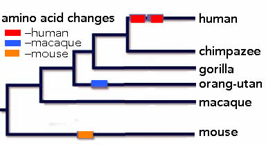
these two amio acid changes alter the activity of the human protein, that is the ensemble of genes that it regulates. That foxp2 has an important role in humans was revealed through studies of individuals in a family that displayed a severe language disorder linked to a mutation that disrupts the function of the foxp2 protein. Individuals carrying this mutant  foxp2 allele display speech apraxia, a “severe impairment in the selection and sequencing of fine oral and facial movements, the ability to break up words into their constituent phonemes, and the production and comprehension of word inflections and syntax” (cited in Bae et al, 2015).  Male mice that carry this foxp2 mutation display changes in the “song” that they sing to female mice (1), while mice carrying a humanized form of foxp2 display changes in “dopamine levels, dendrite morphology, gene expression and synaptic plasticity” in a subset of CNS neurons (2).  While there are many differences between mice and humans, such studies suggest that changes in foxp2 played a role in human evolution, and human speech in particular.

Another way to study the role of human genes using mouse as a model system is to generate what are known as chimeras, named after the creature in Greek mythology composed of parts of multiple organisms.  A couple of years ago, Goldman and colleagues (3) reported that human glial progenitor cells could, when introduced into immune-compromised mice (to circumvent tissue rejection), displaced the mouse’s own glia, replacing them with human glia cells.iPSC transplant 
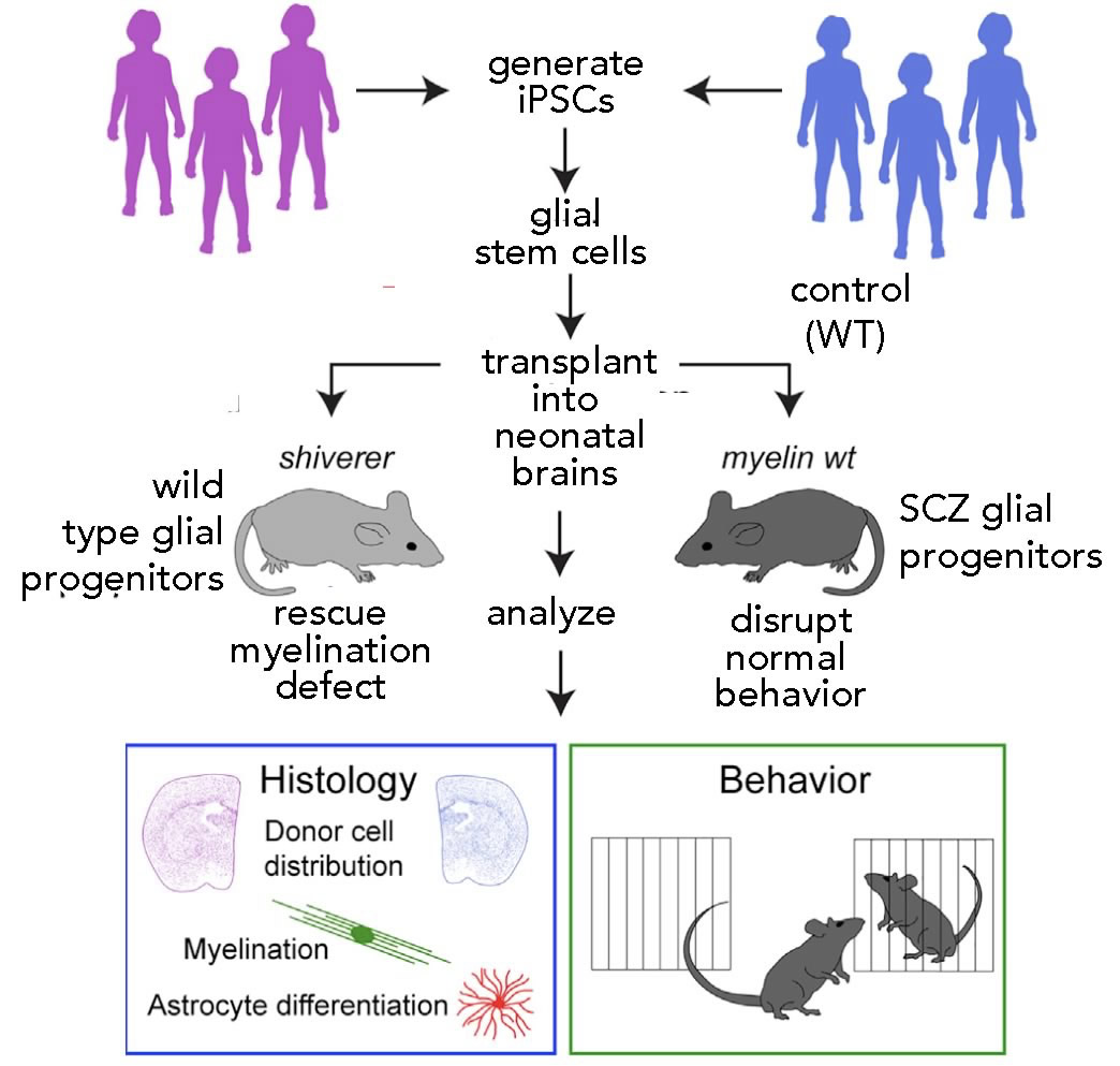
Glial cells are the major non-neuronal component of the central nervous system. Once thought of as passive “support” cells, it is now clear that the two major types of glia, known as astrocytes and oligodendrocytes, play a number of important roles in neural functioning [back track post].  In their early studies, they found that the neurological defects associated with the shaker mutation, a mutation that disrupts the normal behavior of oligodendrocytes, could be rescued by the implantation of normal human glial progenitor cells (hGPCs)(4).  Such studies confirmed what was already known, that the shaker mutation disrupts the normal function of myelin, the insulating structure around axons that dramatically speeds the rate at which neuronal signals (action potentials) move down the axons and activate the links between neurons (synapses). In the central nervous system, myelin is produced by oligodendrocytes as they ensheath neuronal axons.  Human oligodendrocytes derived from hGPCs displaced the mouse’s mutation carrying oligodendrocytes and rescued the shaker mouse’s mutation-associated neurological defect.

golgi staining- diagramSubsequently, Goldman and associates used a variant of this approach to introduce hGPCs (derived from human embryonic stem cells) carrying either a normal or mutant version of the  Huntingtin protein, a protein associated with the severe neural disease Huntington’s chorea (OMIM: 143100)(5).  Their studies strongly support a model that locates defects associated with human Huntington’s disease to defects in glia.  This same research group has generated hGPCs from patient-derived, induced pluripotent stem cells (patient-derived HiPSCs). In this case, the patients had been diagnosed with childhood-onset schizophrenia (SCZ) [link](6).  Skin biopsies were taken from both normal and children diagnosed with SCZ; fibroblasts were isolated, and reprogrammed to form human iPSCs. These iPSCs were treated so that they formed hGPCs that were then injected into mice to generate chimeric (human glial/mouse neuronal) animals. The authors reported systematic differences in the effects of control and SCZ-derived hGPCs; “SCZ glial mice showed reduced prepulse inhibition and abnormal behavior, including excessive anxiety, antisocial traits, and disturbed sleep”, a result that suggests that defects in glial behavior underlie some aspects of the human SCZ phenotype.

The use of human glia chimeric mice provides a powerful research tool for examining the molecular and cellular bases for a subset of human neurological disorders.  Does it raise a question of making mice more human?  Not for me, but perhaps I do not appreciate the more subtle philosophical and ethical issues involved. The mice are still clearly mice, most of their nervous systems are composed of mouse cells, and the overall morphology, size, composition, and organization of their central nervous systems are mouse-derived and mouse-like. The situation becomes rather more complex and potentially therapeutically useful when one talks about generating different types of chimeric animals or of using newly developed genetic engineering tools (the CRISPR CAS9 system found in prokaryotes), that greatly simplify and improve the specificity of the targeted manipulation of specific genes (link).  In these studies the animal of choice is not mice, but pigs – which because of their larger size produce organs for transplantion that are similar in size to the organs of people (see link).  While similar in size, there are two issues that complicate pig to human organ transplantation: first there is the human immune system mediated rejection of foreign  tissue and second there is the possibility that transplantation of porcine organs will lead to the infection of the human recipient with porcine retroviruses.

The issue of rejection (pig into human), always a serious problem, is further exacerbated by the presence in pigs of a gene encoding the enzyme α-1,3 galactosyl transferase (GGTA1). GGTA1 catalyzes the addition of the gal-epitope to a number of cell surface proteins. The gal-epitope is “expressed on the tissues of all mammals except humans and subhuman primates, which have antibodies against the epitope” (7). The result is that pig organs provoke an extremely strong immune (rejection) response in humans.  The obvious technical fix to this (and related problems) is to remove the gal-epitope from pig cells by deleting the GGTA1 enzyme (see 8). It is worth noting that “organs from genetically engineered animals have enjoyed markedly improved survivals in non-human primates” (see Sachs & Gall, 2009).

pig to humanThe second obstacle to pig → human transplantation is the presence of retroviruses within the pig genome.  All vertebrate genomes, including those of humans, contain many inserted retroviruses; almost 50% of the human genome is retrovirus-derived sequence (an example of unintelligent design if ever there was one). Most of these endogenous retroviruses are “under control” and are normally benign (see 9). The concern, however, is that the retroviruses present in pig cells could be activated when introduced into humans. To remove (or minimize) this possibility, Niu et al set out to use the CRISPR CAS9 system to delete these porcine endogenous retroviral sequences (PERVs) from the pig genome; they appear to have succeeded, generating a number of genetically modified pigs without PERVs (see 10).  The hope is that organs generated from PERV-minus pigs from which antigen-generating genes, such as α-1,3 galactosyl transferase, have also been removed or inactivated together with more sophisticated inhibitors of tissue rejection, will lead to an essentially unlimited supply of pig organs that can be used for heart and other organ transplantation (see 11), and so alleviate the delays in transplantation, and so avoid deaths in sick people and the often brutal and criminal harvesting of organs carried out in some countries.

The final strategy being explored is to use genetically modified hosts and patient derived iPSCs  to generate fully patient compatible human organs. To date, pilot studies have been carried out, apparently successfully, using rat embryos with mouse stem cells (see 12 and 13), with much more preliminary studies using pig embryos and human iPSCs (see 14).  The approach involves what is known as chimeric  embryos.  In this case, host animals are genetically modified so that they cannot generate the organ of choice. Typically this is done by mutating a key gene that encodes a transcription factor directly involved in formation of the organ; embryos missing pancreas, kidney, heart, human pig embryo chimera
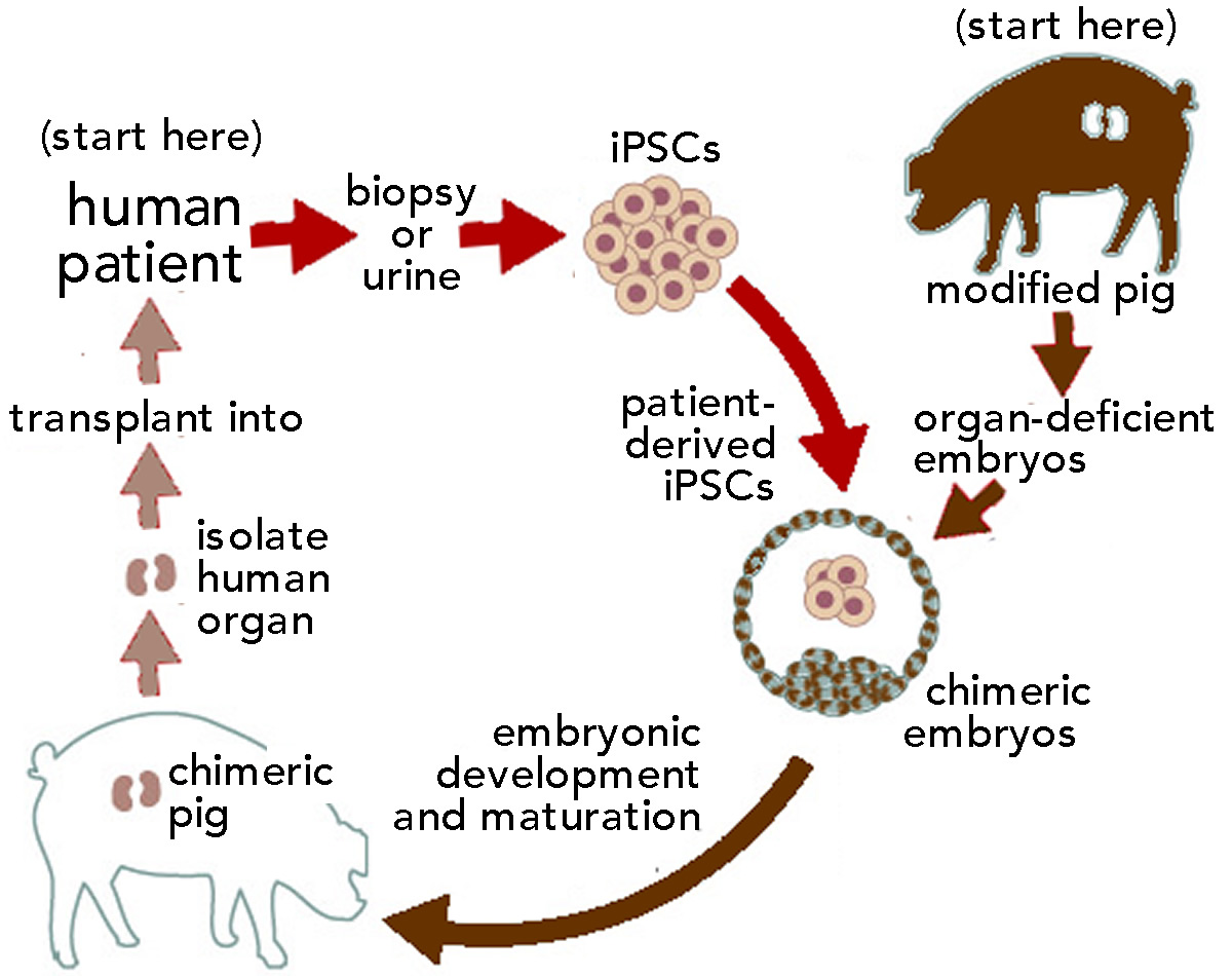
or eyes can be generated.  In an embryo that cannot make these organs, which can be a lethal defect, the introduction of stem cells from an animal that can form these organs can lead to the formation of an organ composed primarily of cells derived from the transplanted (human) cells.

At this point the strategy appears to work reasonably well for mouse-rat chimeras, which are much more closely related, evolutionarily, than are humans and pigs. Early studies on pig-human chimeras appear to be dramatically less efficient. At this point, Jun Wu has been reported as saying of human-pig chimeras that “we estimate [each had] about one in 100,000 human cells” (see 15), with the rest being pig cells.  The bottom line appears to be that there are many technical hurdles to over-come before this method of developing patient-compatible human organs becomes feasible.  Closer to reality are PERV-free/gal-antigen free pig-derived, human compatible organs. The reception of such life-saving organs by the general public, not to mention religious and philosophical groups that reject the consumption of animals in general, or pigs in particular, remains to be seen.

figures reinserted & minor edits 23 October 2020 – new link 17 December 2020.
references cited

  1. A Foxp2 Mutation Implicated in Human Speech Deficits Alters Sequencing of Ultrasonic Vocalizations in Adult Male Mice.
  2. A Humanized Version of Foxp2 Affects Cortico-Basal Ganglia Circuits in Mice
  3. Modeling cognition and disease using human glial chimeric mice.
  4. Human iPSC-derived oligodendrocyte progenitor cells can myelinate and rescue a mouse model of congenital hypomyelination.
  5. Human glia can both induce and rescue aspects of disease phenotype in Huntington disease
  6. Human iPSC Glial Mouse Chimeras Reveal Glial Contributions to Schizophrenia.
  7.  The potential advantages of transplanting organs from pig to man: A transplant Surgeon’s view
  8. see Sachs and Gall. 2009. Genetic manipulation in pigs. and Fisher et al., 2016. Efficient production of multi-modified pigs for xenotransplantation by ‘combineering’, gene stacking and gene editing
  9. Hurst & Magiokins. 2017. Epigenetic Control of Human Endogenous Retrovirus Expression: Focus on Regulation of Long-Terminal Repeats (LTRs)
  10. Nui et al., 2017. Inactivation of porcine endogenous retrovirus in pigs using CRISPR-Cas9
  11. Zhang  2017. Genetically Engineering Pigs to Grow Organs for People
  12. Kobayashi et al., 2010. Generation of rat pancreas in mouse by interspecific blastocyst injection of pluripotent stem cells.
  13. Kobayashi et al., 2015. Targeted organ generation using Mixl1-inducible mouse pluripotent stem cells in blastocyst complementation.
  14. Wu et al., 2017. Interspecies Chimerism with Mammalian Pluripotent Stem Cells
  15. Human-Pig Hybrid Created in the Lab—Here Are the Facts

Reverse Dunning-Kruger effects and science education

The Dunning-Kruger (DK) effect is the well-established phenomenon that people tend to over estimate their understanding of a particular topic or their skill at a particular task, often to a dramatic degree [link][link]. We see examples of the DK effect throughout society; the current administration (unfortunately) and the nutritional supplements / homeopathy section of Whole Foods spring to mind as examples. But there is a less well-recognized “reverse DK” effect, namely the tendency of instructors, and a range of other public communicators, to over-estimate what the people they are talking to are prepared to understand, appreciate, and accurately apply. The efforts of science communicators and instructors can be entertaining but the failure to recognize and address the reverse DK effect results in ineffective educational efforts. These efforts can themselves help generate the illusion of understanding in students and the broader public (discussed here). While a confused understanding of the intricacies of cosmology or particle physics can be relatively harmless in their social and personal implications, similar misunderstandings become personally and publicly significant when topics such as vaccination, alternative medical treatments, and climate change are in play.

There are two synergistic aspects to the reverse DK effect that directly impact science instruction: the need to understand what one’s audience does not understand together with the need to clearly articulate the conceptual underpinnings needed to understand the subject to be taught. This is in part because modern science has, at its core, become increasingly counter-intuitive over the last approximately 100 years or so, a situation that can cause serious confusions that educators must address directly and explicitly. The first reverse DK effect involves the extent to which the instructor (and by implication the course and textbook designer) has an accurate appreciation of what students think or think they know, what ideas they have previously been exposed to, and what they actually understand about the implications of those ideas.  Are they prepared to learn a subject or does the instructor first have to acknowledge and address conceptual confusions and build or rebuild base concepts?  While the best way to discover what students think is arguably a Socratic discussion, this only rarely occurs for a range of practical reasons. In its place, a number of concept inventory-type testing instruments have been generated to reveal whether various pre-identified common confusions exist in students’ thinking. Knowing the results of such assessments BEFORE instruction can help customize how the instructor structures the learning environment and content to be presented and whether the instructor gives students the space to work with these ideas to develop a more accurate and nuanced understanding of a topic.  Of course, this implies that instructors have the flexibility to adjust the pace and focus of their classroom activities. Do they take the time needed to address student issues or do they feel pressured to plow through the prescribed course content, come hell, high water, or cascading student befuddlement.

A complementary aspect of the reverse DK effect, well-illustrated in the “why magnets attract” interview with the physicist Richard Feynman, is that the instructor, course designer, or textbook author(s) needs to have a deep and accurate appreciation of the underlying core knowledge necessary to understand the topic they are teaching. Such a robust conceptual understanding makes it possible to convey the complexities involved in a particular process and explicitly values appreciating a topic rather than memorizing it.  It focuses on the general, rather than the idiosyncratic. A classic example from many an introductory biology course is the difference between expecting students to remember the steps in glycolysis or the Krebs cycle reaction system, as opposed to the general principles that underlie the non-equilibrium reaction networks involved in all biological functions, a reaction network based on coupled chemical reactions and governed by the behaviors of thermodynamically favorable and unfavorable reactions. Without a explicit discussion of these topics, all too often students are required to memorize names without understanding the underlying rationale driving the processes involved; that is, why the system behaves as it does.  Instructors also give false “rubber band” analogies or heuristics to explain complex phenomena (see Feynman video 6:18 minutes in). A similar situation occurs when considering how molecules come to associate and dissociate from one another, for example in the process of regulating gene expression or repairing mutations in DNA. Most textbooks simply do not discuss the physiochemical processes involved in binding specificity, association, and dissociation rates, such as the energy changes associated with molecular interactions and thermal collisions (don’t believe me? look for yourself!). But these factors are essential for a student to understand the dynamics of gene expression [link], as well as the specificity of modern methods involved in genetic engineering, such as restriction enzymes, polymerase chain reaction, and CRISPR CAS9-mediated mutagenesis. By focusing on the underlying processes involved we can avoid their trivialization and enable students to apply basic principles to a broad range of situations. We can understand exactly why CRISPR CAS9-directed mutagenesis can be targeted to a single site within a multibillion-base pair genome.

Of course, as in the case of recognizing and responding to student misunderstandings and knowledge gaps, a thoughtful consideration of underlying processes takes course time, time that trades the development of a working understanding of core processes and principles for broader “coverage” of frequently disconnected facts, the memorization and regurgitation of which has been privileged over understanding why those facts are worth knowing. If our goal is for students to emerge from a course with an accurate understanding of the basic processes involved rather than a superficial familiarity with a plethora of unrelated facts, however, a Socratic interaction with the topic is essential. What assumptions are being made, where do they come from, how do they constrain the system, and what are their implications?  Do we understand why the system behaves the way it does? In this light, it is a serious educational mystery that many molecular biology / biochemistry curricula fail to introduce students to the range of selective and non-selective evolutionary mechanisms (including social and sexual selection – see link), that is, the processes that have shaped modern organisms.

Both aspects of the reverse DK effect impact educational outcomes. Overcoming the reverse DK effect depends on educational institutions committing to effective and engaging course design, measured in terms of retention, time to degree, and a robust inquiry into actual student learning. Such an institutional dedication to effective course design and delivery is necessary to empower instructors and course designers. These individuals bring a deep understanding of the topics taught and their conceptual foundations and historic development to their students AND must have the flexibility and authority to alter the pace (and design) of a course or a curriculum when they discover that their students lack the pre-existing expertise necessary for learning or that the course materials (textbooks) do not present or emphasize necessary ideas. Radiation-kills-in-BoulderUnfortunately, all too often instructors, particularly in introductory level college science courses, are not the masters of their ships; that is, they are not rewarded for generating more effective course materials. An emphasis on course “coverage” over learning, whether through peer-pressure, institutional apathy, or both, generates unnecessary obstacles to both student engagement and content mastery.  To reverse the effects of the reverse DK effect, we need to encourage instructors, course designers, and departments to see the presentation of core disciplinary observations and concepts as the intellectually challenging and valuable endeavor that it is. In its absence, there are serious (and growing) pressures to trivialize or obscure the educational experience – leading to the socially- and personally-damaging growth of fake knowledge.

empty images holders removed, new image added – 17 December 2020

Is it time to start worrying about conscious human “mini-brains”?

A human iPSC cerebral organoid in which pigmented retinal epithelial cells can be seen (from the work of McClure-Begley et al.).   Also see “Can lab-grown brains become conscious?” by Sara Readon Nature 2020.

The fact that experiments on people are severely constrained is a major obstacle in understanding human development and disease.  Some of these constraints are moral and ethical and clearly appropriate and necessary given the depressing history of medical atrocities.  Others are technical, associated with the slow pace of human development. The combination of moral and technical factors has driven experimental biologists to explore the behavior of a wide range of “model systems” from bacteria, yeasts, fruit flies, and worms to fish, frogs, birds, rodents, and primates.  Justified by the deep evolutionary continuity between these organisms (after all, all organisms appear to be descended from a single common ancestor and share many molecular features), experimental evolution-based studies of model systems have led to many therapeutically valuable insights in humans – something that I suspect a devotee of intelligent design creationism would be hard pressed to predict or explain (post link).

While humans are closely related to other mammals, it is immediately obvious that there are important differences – after all people are instantly recognizable from members of other closely related species and certainly look and behave differently from mice. For example, the surface layer of our brains is extensively folded (they are known as gyrencephalic) while the brain of a mouse is smooth as a baby’s bottom (and referred to as lissencephalic). In humans, the failure of the brain cortex to fold is known as lissencephaly, a disorder associated with severe neurological defects. With the advent of more and more genomic sequence data, we can identify human specific molecular (genomic) differences. Many of these sequence differences occur in regions of our DNA that regulate when and where specific genes are expressed.  Sholtis & Noonan (1) provide an example: the HACNS1 locus is a 81 basepair region that is highly conserved in various vertebrates from birds to chimpanzees; there are 13 human specific changes in this sequence that appear to alter its activity, leading to human-specific changes in the expression of nearby genes (↓). At this point ~1000 genetic elements that are different in humans compared to other vertebrates have been identified and more are likely to emerge (2).  Such human-specific changes can make modeling human-specific behaviors, at the cellular, tissue, organ, and organism level, in non-human model systems difficult and problematic (3, 4).   It is for this reason that scientists have attempted to generate better human specific systems.

human sequence divergence

One particularly promising approach is based on what are known as embryonic stem cells (ESCs) or pluripotent stem cells (PSCs). Human embryonic stem cells are generated from the inner cell mass of a human embryo and so involve the destruction of that embryo – which raises a number of ethical and religious concerns as to when “life begins” (5).  Human pluripotent stem cells are isolated from adult tissues but in most cases require invasive harvesting methods that limit their usefulness.  Both ESCs and PSCs can be grown in the laboratory and can be induced to differentiate into what are known as gastruloids.  Such gastruloids can develop anterior-posterior (head-tail), dorsal-ventral (back-belly), and left-right axes analogous to those found in embryos (6) and adults (top panel ↓). In the case of PSCs, the gastruloid (bottom panel ↓) is essentially a twin of the organism from which the PSCs were derived, a situation that raises difficult questions: is it a distinct individual, is it the property of the donor or the creation of a technician.  The situation will be further complicated if (or rather, when) it becomes possible to generate viable embryos from such gastruloids.

Axes

gastruloid-embryo-comparisonThe Nobel prize winning work of Kazutoshi Takahashi and Shinya Yamanaka (7), who devised methods to take differentiated (somatic) human cells and reprogram them into ESC/PSC-like cells, cells known as induced pluripotent stem cells (iPSCs)(8), represented a technical breakthrough that jump-started this field. While the original methods derived sample cells from tissue biopsies, it is possible to reprogram kidney epithelial cells recovered from urine, a non-invasive approach (910).  Subsequently, Madeline Lancaster, Jurgen Knōblich, and colleagues devised an approach by which such cells could be induced to form what they termed “cerebral organoids” (although Yoshiki Sasai and colleagues were the first to generate neuronal organoids); they used this method to examine the developmental defects associated with microencephaly (11).  The value of the approach was rapidly recognized and a number of studies on human conditions, including  lissencephaly (12), Zika-virus infection-induced microencephaly (13), and Down’s syndrome (14);  investigators have begun to exploit these methods to study a range of human diseases – and rapid technological progress is being made.

The production of cerebral organoids from reprogrammed human somatic cells has also attracted the attention of the media (15).  While “mini-brain” is certainly a catchier name, it is a less accurate description of a cerebral organoid, itself possibly a bit of an overstatement, since it is not clear exactly how “cerebral” such organoids are. For example, the developing brain is patterned by embryonic signals that establish its asymmetries; it forms at the anterior end of the neural tube (the nascent central nervous system and spinal cord) and with distinctive anterior-posterior, dorsal-ventral, and left-right asymmetries, something that simple cerebral organoids do not display.  Moreover, current methods for generating cerebral organoids involve primarily what are known as neuroectodermal cells – our nervous system (and that of other vertebrates) is a specialized form of the embryo’s surface layer that gets internalized during development. In the embryo, the developing neuroectoderm interacts with cells of the circulatory system (capillaries, veins, and arteries), formed by endothelial cells and what are known as pericytes that surround them. These cells, together with interactions with glial cells (astrocytes, a non-neuronal cell type) combine to form the blood brain barrier.  Other glial cells (oligodendrocytes) are also present; in contrast, both types of glia (astrocytes and oligodendrocytes) are rare in the current generation of cerebral organoids. Finally, there are microglial cells,  immune system cells that originate from outside the neuroectoderm; they invade and interact with neurons and glia as part of the brain’s dynamic neural capillary and neuronssystem. The left panel of the figure shows, in highly schematic form how these cells interact (16). The right panel is a drawing of neural tissue stained by the Golgi method (17), which reveals ~3-5% of the neurons present. There are at least as many glial cells present, as well as microglia, none of which are visible in the image. At this point, cerebral organoids typically contain few astrocytes and oligodendrocytes, no vasculature, and no microglia. Moreover, they grow to be about 1 to 3 mm in diameter over the course of 6 to 9 months; that is significantly smaller in volume than a fetal or newborn’s brain. While cerebral organoids can generate structures characteristic of retinal pigment epithelia (top figure) and photo-responsive neurons (18), such as those associated with the retina, an extension of the brain, it is not at all clear that there is any significant sensory input into the neuronal networks that are formed within a cerebral organoid, or any significant outputs, at least compared to the role that the human brain plays in controlling bodily and mental functions.

The reasonable question, then, must be whether a  cerebral organoid, which is a relatively simple system of cells (although itself complex), is conscious. It becomes more reasonable as increasingly complex systems are developed, and such work is proceeding apace. Already researchers are manipulating the developing organoid’s environment to facilitate axis formation, and one can anticipate the introduction of vasculature. Indeed, the generation of microglia-like cells from iPSCs has been reported; such cells can be incorporated into cerebral organoids where they appear to respond to neuronal damage in much the same way as microglia behave in intact neural tissue (19).

We can ask ourselves, what would convince us that a cerebral organoid, living within a laboratory incubator, was conscious? How would such consciousness manifest itself? Through some specific pattern of neural activity, perhaps?  As a biologist, albeit one primarily interested in molecular and cellular systems, I discount the idea, proposed by some physicists and philosophers as well as the more mystical, that consciousness is a universal property of matter (20,21).  I take consciousness to be an emergent property of complex neural systems, generated by evolutionary mechanisms, built during embryonic and subsequent development, and influenced by social interactions (BLOG LINK) using information encoded within the human genome (something similar to this: A New Theory Explains How Consciousness Evolved). While a future concern, in a world full of more immediate and pressing issues, it will be interesting to listen to the academic, social, and political debate on what to do with mini-brains as they grow in complexity and perhaps inevitably, towards consciousness.

Footnotes and references

Thanks to Rebecca Klymkowsky, Esq. and Joshua Sanes, Ph.D. for editing and disciplinary support. Minor updates and the reintroduction of figures 22 Oct. 2020.

  1. Gene regulation and the origins of human biological uniqueness
  2.  See also Human-specific loss of regulatory DNA and the evolution of human-specific traits
  3. The mouse trap
  4. Mice Fall Short as Test Subjects for Some of Humans’ Deadly Ill
  5. The status of the human embryo in various religions
  6. Interactions between Nodal and Wnt signalling Drive Robust Symmetry Breaking and Axial Organisation in Gastruloids (Embryonic Organoids)
  7.  Induction of pluripotent stem cells from mouse embryonic and adult fibroblast cultures by defined factors
  8.  How iPS cells changed the world
  9.  Generation of Induced Pluripotent Stem Cells from Urine
  10. Urine-derived induced pluripotent stem cells as a modeling tool to study rare human diseases
  11. Cerebral organoids model human brain development and microcephaly.
  12. Human iPSC-Derived Cerebral Organoids Model Cellular Features of Lissencephaly and Reveal Prolonged Mitosis of Outer Radial Glia
  13. Using brain organoids to understand Zika virus-induced microcephaly
  14. Probing Down Syndrome with Mini Brains
  15. As an example, see The Beauty of “Mini Brains”
  16. Derived from Central nervous system pericytes in health and disease
  17. Golgi’s method .
  18. Cell diversity and network dynamics in photosensitive human brain organoids
  19. Efficient derivation of microglia-like cells from human pluripotent stem cells
  20. The strange link between the human mind and quantum physics – BBC:
  21. Can Quantum Physics Explain Consciousness?